Fluorescence microscopy is an imaging technique that uses fluorescence to generate an image, allowing for the visualization of specific structures within a sample. Unlike classical microscopy, which relies on light absorption, fluorescence microscopy can highlight particular components by tagging them with fluorescent dyes or proteins. This method provides higher specificity and sensitivity, enabling researchers to observe the dynamics of live cells and molecular interactions in real-time. Additionally, it allows for multi-color labeling, which can distinguish between multiple targets within a single specimen.
Multiphoton microscopy, such as two-photon excitation (TPE) microscopy, uses multiple low-energy photons to excite a fluorophore, allowing for deeper tissue penetration with reduced phototoxicity. This technique is particularly advantageous for imaging live tissues and thick specimens, as it minimizes damage and provides high-resolution, three-dimensional images.
The most demanding applications require precisely adjusted excitation laser parameters to enhance image quality. Our product portfolio includes lasers tailored specifically for multiphoton microscopy, e.g., the ModeSHG780 with excellent stability, and pulse quality, ensuring your research achieves the highest level of detail and accuracy. Trusted by leading scientists worldwide, our products set the standard for excellence in multiphoton microscopy, making us the top choice for your advanced imaging needs.
RELATED PRODUCTS
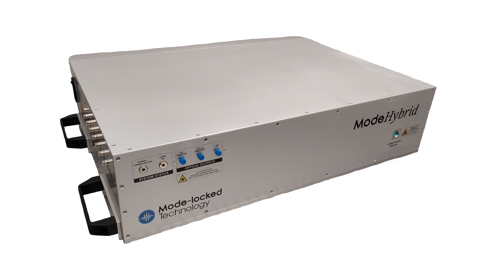
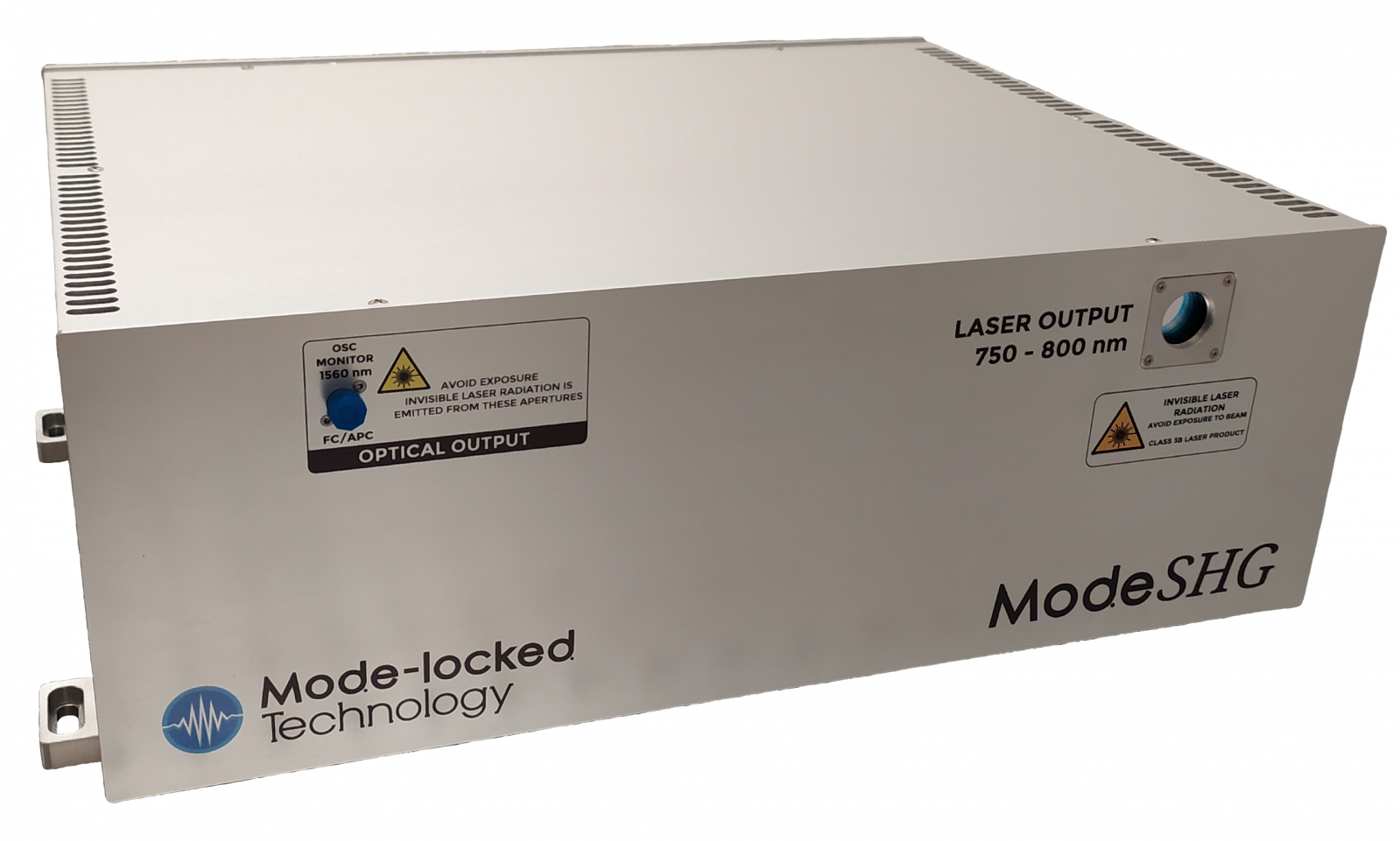 ModeSHG780 ModeSHG780 |
|
ModeLDC |